


| Received: October 8 2023; Accepted: November 23 2023 |
Autoimmune anti-NMDAR encephalitis is a serious, underrecognized, rapidly progressive, and potentially fatal yet treatable condition whose diagnostic criteria were defined in 2016.1,2 Up to 90% of cases begin with psychiatric symptoms, and 77% are initially evaluated by psychiatrists, making it challenging to distinguish from a primary psychiatric disorder.2,3 This correspondence aims to emphasize anti-NMDAR encephalitis as a differential diagnosis in patients with subacute psychiatric manifestations.
We present two clinical cases treated at university general hospitals in Rio Grande do Sul, Brazil, followed by an integrative literature review in the PubMed and SciELO databases, conducted in 2020 and 2023. In both cases, the patient or legal guardian provided written informed consent.
The first case, a 25 year-old woman, was admitted to a psychiatric unit due to mutism and catatonia; the second, an 11 year-old boy, was admitted to a pediatrics unit for suicide risk and aggression towards others. Over a period of 4 or more weeks, they both developed neuropsychiatric alterations after initial depressive symptomatology linked to recent family losses. Their neuropsychiatric presentation included disorganized behavior with recurrent agitation, hypersexualization, psychosis, unprovoked laughter, aggression towards others, as well as cognitive changes, orofacial movement disorders, speech disturbances, impaired writing, and progressive decline in self-care and autonomy. In terms of general clinical symptoms, the female patient experienced tonic-clonic epileptic seizures and dysautonomia, while the male patient presented with flu-like symptoms at onset. Neither had a history of psychiatric disorders, and typical findings were absent in complementary exams (i.e., electroencephalogram, blood work, and magnetic resonance imaging were not suggestive) (Box 1). The diagnosis was confirmed through indirect immunofluorescence by the presence of anti-NMDAR neuronal antibodies in cerebrospinal fluid, the only specific diagnostic test for this condition.4
| Additional tests | Case 1 | Case 2 |
|---|---|---|
| Laboratory |
|
|
| Cerebrospinal fluid |
|
|
| ECG and EEG |
|
|
| Imaging tests |
|
|
AFP = alpha-fetoprotein; ANA = antinuclear antibody; anti-HCV = hepatitis C antibody test; anti-NMDAR = anti-N-methyl-D-aspartate receptor; HCG = human chorionic gonadotropin; CA 125 = cancer antigen 125; CRP = C-reactive protein; CT = computed tomography; ECG = electrocardiogram; EEG = electroencephalogram; ESR = erythrocyte sedimentation rate; HbsAg = hepatitis B surface antigen; LDH = lactate dehydrogenase; MRI = magnetic resonance imaging; PCR = polymerase chain reaction; RF = rheumatoid factor; TgAb = thyroglobulin antibodies; TPO = thyroid peroxidase; TRAb = thyrotropin receptor antibody; VDRL = venereal disease research laboratory.
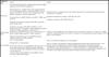
Nearly half of autoimmune psychosis patients may have a pre-existing psychiatric disorder, most commonly depression.2 The most common psychiatric symptoms of anti-NMDAR encephalitis are agitation (59%), psychosis (54%), catatonia (42% in adults and 35% in children) and mood disorders (27%).2 In 4% of cases, psychiatric symptoms may be the sole presentation.3 It is estimated that 37% of cases start before 18 years of age, with 70% showing prodromal flu-like symptoms in the two weeks before onset of encephalitis manifestations.3,4,5 Movement disorders occur in 75% of adults and 95% of children.3 As shown in this case report, a lack of typical findings in complementary exams does not rule out diagnosis, although up to 80% of cases show cerebrospinal fluid changes (moderate lymphocytic pleocytosis) and more than 90% show electroencephalogram changes.2,4
However, cranial magnetic resonance imaging can be normal in up to 50-70% of cases or reveal non-specific hyperintense lesions in up to 50% of later-stage cases and 35% of early-stage cases.2,4 Electroconvulsive therapy is recognized as an effective and safe first-line treatment for malignant catatonia, leading to complete (60%) or partial (33%) neurological recovery in these patients.4,6 It also enhances the effectiveness of treatment with immunomodulators, allowing complete remission of psychiatric symptoms and associated risks.4,6
ACKNOWLEDGMENTS
The authors would like to thank our colleagues, Pedro Henrique Manfro and Manoela Prevedello Ceretta, as well as Liselotte Menke Barea MD, PhD, associate professor of neurology at the Universidade Federal de Ciências da Saúde de Porto Alegre (UFCSPA), coordinator of the research center (PESQUINEURO) and head of the neurology service at the Irmandade Santa Casa de Misericórdia de Porto Alegre. They were involved in the assessment, discussion, and description of the clinical cases reported in this study. Their contributions were crucial for the accurate diagnosis, treatment, and subsequent favorable outcomes in both patients.
DISCLOSURE
The authors report no conflicts of interest.
 PDF
 Print
 Send this article by email
 How to cite this article
 Submit a comment
 Mendeley
 Pocket
 Share on Facebook
 Share on Twitter