


ABSTRACT:
OBJECTIVE: Galectins (Gal), which have been linked with inflammatory response in the central
nervous system, may play an important role in the pathogenesis of bipolar disorder.
In this study, we investigated whether serum Gal-1 and Gal-3 levels are related to
bipolar disorder.
METHODS: Thirty-six patients diagnosed with bipolar disorder were included. C-reactive protein,
Gal-1, Gal-3 serum concentrations were evaluated on the first day of hospitalization
and the third week of treatment and were compared with 41 healthy controls. Illness
severity was evaluated with the Young Mania Rating Scale.
RESULTS: Upon hospitalization, the C-reactive protein levels of bipolar disorder patients were
significantly higher than in the third week of treatment or in healthy controls. Gal-1
levels on the first day of hospitalization and the third week of treatment were higher
than those of healthy controls.There was no significant difference between patient
Gal-3 levels upon hospitalization and those of healthy controls; at the end of the
third week of treatment, Gal-3 levels were significantly higher than on the first
day of hospitalization.
CONCLUSION: Our study is the first to show a change in Gal levels after treatment and to evaluate
the role of Gal in bipolar disorder. Gal-1 may play a role in the pathophysiology
of bipolar disorder. Gal-3 could be a biomarker candidate for assessing treatment
response.
Keywords: Galectins; bipolar disorder; inflammation; mood disorders; biomarkers.
FIGURES
| Received: January 1 2024; Accepted: April 23 2024 |
INTRODUCTION
Bipolar disorder (BD), a serious mental disorder with a lifetime prevalence of 1.3-5.0%, is characterized by recurrent manic and depressive episodes.1,2 Although its etiology and pathogenesis are still unclear, it is known that genetic, environmental, neurobiological, hormonal, and neurochemical factors are involved.3 Inflammatory processes are a relatively novel and important area of research on the etiological and pathophysiological factors of BD.4 Inflammation refers to a series of complex changes that occur in response to tissue damage caused by various factors (e.g., microorganisms, toxic substances, or trauma), including the activation and recruitment of immune cells, increased blood flow, and vascular permeability.5
Abnormalities in peripheral inflammatory markers in patients with BD have been widely reported in previous case-control studies,6,7 and many studies have observed higher serum concentrations of inflammatory cytokines in patients with BD.8-11 Neuroimmune response involves innate immune reactions in the central nervous system (CNS), often associated with chemically induced damage.12 Research into the etiology of BD primarily focuses on various factors, such as the immune system, neuroimaging, genetics, blood biochemistry, the endocrine system, and biological rhythms. The emergence of neuroimmune response to peripheral inflammatory changes in BD has become a significant area of research on the origins and progression of the disease. A growing body of evidence indicates that the alterations observed in the CNS of BD patients may stem from immune system activation and the release of inflammatory substances. Investigations into changes in pro-inflammatory (interleukin-6 and tumor necrosis factor-a) and anti-inflammatory cytokines in both the CNS and peripheral blood of BD patients have shown consistent results.13,14 It is now recognized that the neuroimmune system plays a crucial role in the pathological mechanisms of BD, and biomarkers associated with neuroinflammatory responses are an important focus of research. The available evidence indicates that inflammatory response differs between remission and BD episodes and that there is a relationship between varying cytokine levels and these episodes.15
Various proteins have been investigated as candidate biomarkers in the relationship between inflammation and mental disorders. One of these is galectin (Gal), a class of proteins belonging to the ß-galactocyte-binding lectin family. Gal plays important roles in the regulation of immune and inflammatory responses.3 These proteins are found both inside and outside of cells. Extracellular Gal interacts with glycoproteins on the cell surface and in the extracellular matrix, while intracellular Gal interacts with cytoplasmic and nuclear proteins in a carbohydrate-independent manner.16-18 They are involved in a variety of biological processes, including cell adhesion, migration, proliferation, transformation, apoptosis, angiogenesis, and immune response.19
Members of the Gal family play different roles in mediating pathological processes in a variety of inflammatory illnesses.20 Under normal physiological circumstances, Gal plays an important role in CNS balance through processes such as neuronal myelination, proliferation of neuronal stem cells, and transportation of apical vesicles within neuronal cells.21 Consequently, Gal serves as a significant modulator in CNS equilibrium and neuroinflammation. In models of neuroinflammatory disease, Gal can act either as an extracellular mediator or an intracellular regulator, influencing inflammatory reaction or facilitating tissue remodeling in damaged CNS regions.3 There's a growing belief that Gal could be implicated in neuropsychiatric disorders, particularly neurodegeneration and the associated neuroimmune response.
Gal-1 was the first protein discovered in the Gal family. The structure of human Gal-1 includes a six-strand, five-arm ß-sheet in an anti-parallel arrangement.22 Gal-1 is expressed in a variety of tissues (e.g. liver, spleen, lung, brain, and skeletal muscle) and is found in the cytosol and extracellular space.23,24
Gal-3 is the only member of the chimera-type Gal group. It affects the differentiation and growth of various immune cells, inducing apoptosis in T cells and neutrophils. T cells activate a variety of myeloid and lymphoid cells that can cause mediator release and cytokine production (e.g., monocytes, mast cells, and neutrophils).21,25 Gal-3 is also expressed in inflammatory cells, including astrocytes, microglia, macrophages, dendritic cells, eosinophils, mast cells, natural killer cells, and activated T and B cells.26 So far, studies have shown that Gal plays an important role in immune and inflammatory response. Several studies have proven the key roles of Gal-1 and Gal-3 in neuroimmune response to various neuropathological conditions.27 Kajitani et al.28 reported that the Gal-1 levels of patients with schizophrenia did not differ significantly from healthy controls, and their Gal-3 levels were high. Yüksel et al.29 examined the relationship between serum Gal-1 levels in patients with schizophrenia, healthy siblings of patients with schizophrenia, and a healthy control group, reporting that there were no significant differences in Gal-1 and Gal-3 levels between healthy controls and patients with schizophrenia. Another study reported that patients with schizophrenia had lower Gal-3 levels than healthy controls.30 In relatives of patients with schizophrenia, Gal-1 and Gal-3 are candidate biomarkers for protection against genetic burden and illness development. To the best of our knowledge, no research has been conducted on Gal in patients diagnosed with BD.
The purpose of this study was to determine whether Gal plays a role in the pathogenesis of BD. The Gal-1 and Gal-3 levels of patients with BD, assessed upon hospitalization for a manic episode and after the third week of treatment, were compared with healthy controls.
METHODS
Hospitalized patients aged 18-65 years who had been evaluated by an experienced psychiatrist and diagnosed with a BD manic episode according to the DSM-5 and were referred to Ankara City Hospital Psychiatry Clinic were included in the study. Healthy controls were selected among willing hospital staff who were matched to patients by age and sex. None of the staff had been diagnosised or treated for a mental disorder. Considering that inflammatory processes would be affected, anyone with the following conditions was excluded from the study: pregnancy, morbid obesity (body mass index > 40), systemic inflammatory disease, mental retardation, or neurocognitive disorders. The participants were included in the study after undergoing evaluation by an experienced psychiatrist with the Structured Clinical Interview for DSM-5.
A total of 36 patients who met DSM-5 criteria for manic episodes and 41 healthy controls were included in the study. Seven of the patients were having a first episode. Of the remaining 29, 21 had stopped taking their medication in the month prior to admission and 8 were using psychotropics. On the first day of hospitalization, the patients' psychometric evaluation was performed and their blood samples were taken. These procedures were repeated for all 30 inpatients at the end of the third week of treatment. The in-hospital drug treatments were recorded. A total of 30 patients who scored < 5 in the Young Mania Rating Scale (YMRS) and < 7 in the Hamilton Depression Rating Scale were considered to be in remission.
Young Mania Rating Scale
The development of scales to measure the severity of manic episodes began in the 1970s, with Young et al. publishing the YMRS in 1978. The YMRS, the most widely used scale for measuring manic state severity in clinical studies, consists of 11 items, seven of which use a 5-point Likert scale and four of which use a 9-point scale. Scores range from 0 to 44 points. The validity and reliability of the Turkish version were tested by Karadag et al.31
Hamilton Depression Rating Scale
The Hamilton Depression Rating Scale, the most widely used instrument to assess depression severity, is not used for diagnosis but to evaluate treatment response. The original scale, released in 1960, included 17 items that evaluated 17 depression symptoms in three or five dimensions. Scores of 16-25, 26-40, and = 40 are considered indicative of mild, moderate, and severe depressive syndrome, respectively.32 The validity and reliability of the Turkish version were tested by Akdemir et al.33
Biochemical analysis
The blood samples were taken after a 12-hour fast at 8:00 a.m. via venipuncture into a tube containing BD Vacutainer SST II Advance serum separator gel and were centrifuged at 1,500 g for 10 minutes. They were then transferred to Eppendorf tubes and kept at -80 °C until the day of analysis. The samples were thawed for 12 hours at room temperature before the enzime-linked immunosorbent assay (ELISA) was performed. Serum Gal-1 and Gal-3 levels were measured with ELISA assay kits (Human Galectin 1 ELISA kit, Human Galectin 3 ELISA kit) according to manufacturer instructions. Gal-1 and Gal-3 values were expressed as ng/mL.
Statistical analysis
For continuous numerical variables, the normality of distribution was determined with the Shapiro-Wilk test. Descriptive statistics were expressed as mean (SD) and median (minimum-maximum) for continuous numerical variables, while categorical variables were expressed as the number of cases and percentages.
After the goodness-of-fit tests, Student's t-test was used to determine the significance of the difference in continuous numerical variables that met parametric test assumptions. When the number of independent groups was two, the Mann-Whitney U test was used to determine significance of the difference in terms of continuous numerical variables when parametric test assumptions were not met. Spearman's rank correlation coefficient was used to determine whether the correlation between continuous numerical variables was statistically significant. The Pearson chi-square test or Fisher's exact test was used to compare categorical variables. The Kruskal-Wallis test was used to compare the continuous variables of more than two groups.
The data were analyzed in IBM SPSS Statistics 25.0 and the results were considered statistically significant at p < 0.05.
Ethics statement
This study was approved on December 2, 2020 by the Health Sciences University Ankara City Hospital No. 2 Clinical Research Ethics Committee (decision 20-04).
RESULTS
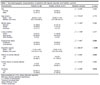
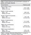
A total of 36 patients diagnosed with a BD manic episode and 41 healthy controls were included in the study (41.7% men, 58.3% women). The mean participant age was 35.50 (SD, 11.54) years. The overall smoking rate was 61.1% and was significantly higher among patients than controls (p = 0.018). The sociodemographic characteristics of the BD and control groups are compared in Table 1. Seven (19.4%) of the patients were having their first episode and had no history of medication, 58.3% had stopped using their medication in the last month, and the mean illness duration was 9.52 (SD, 10.99) years. The patients' clinical information and illness severity are shown in Table 2.
| Characteristics | Patients (n=36) | Healthy controls (n=41) | Statistical analysis | p-value |
|---|---|---|---|---|
| Sex | ?2 = 0.039┼ | 0.843 | ||
| Female | 21 (58.3) | 23 (56.1) | ||
| Male | 15 (41.7) | 18 (43.9) | ||
| Age (years) | U = 710.00╬ | 0.775 | ||
| Mean ± SD | 35.50±11.54 | 35.85±11.29 | ||
| Median (min-max) | 33.0 (18.0-65.0) | 31.0 (20.0-61.0) | ||
| Marital status | ?2 = 1.303§ | 0.560 | ||
| Married | 14 (38.9) | 21 (51.2) | ||
| Single | 18 (50.0) | 17 (41.5) | ||
| Divorced | 4 (11.1) | 3 (7.3) | ||
| Living condition | ?2 = 1.796§ | 0.477 | ||
| Alone | 8 (22.2) | 11 (26.8) | ||
| With family | 28 (50.0) | 28 (50.0) | ||
| With friend(s) | 0 (0.0) | 2 (4.9) | ||
| Employment status | ?2 = 7.967┼ | 0.005 | ||
| Employed | 18 (50.0) | 33 (80.5) | ||
| Unemployed | 18 (50.0) | 8 (19.5) | ||
| Education (years) | U = 398.50╬ | < 0.001 | ||
| Mean ± SD | 11.22±4.40 | 14.56±4.16 | ||
| Median (min-max) | 12.0 (5.0-17.0) | 16.0 (5.0-18.0) | ||
| History of suicide attempt | ?2 = 4.805┼ | 0.043 | ||
| Yes | 4 (11.9) | 0 (0.0) | ||
| No | 32 (88.1) | 41 (100.0) | ||
| Family history of suicide attempt | ?2 = 0.018┼ | 0.990 | ||
| Yes | 2 (5.6) | 2 (4.9) | ||
| No | 34 (94.4) | 39 (95.1) | ||
| Smoking | ?2 = 5.599┼ | 0.018 | ||
| Yes | 22 (61.1) | 14 (34.1) | ||
| No | 14 (38.9) | 27 (65.9) |
| Clinical features | Patients (n=36) |
|---|---|
| Illness duration (years) | |
| Mean ± SD | 9.52±10.99 |
| Median (min-max) | 4.5 (0.1-48.0) |
| Number of manic episodes | |
| Mean ± SD | 3.66±3.32 |
| Median (min-max) | 2.0 (1.0-15.0) |
| Number of depressive episodes | |
| Mean ± SD | 1.00±1.89 |
| Median (min-max) | 0.0 (0.0-10.0) |
| Number of hospitalizations | |
| Mean ± SD | 3.11±2.95 |
| Median (min-max) | 2.0 (0.0-15.0) |
| YMRS (upon hospitalization) | |
| Mean ± SD | 34.50±7.81 |
| Median (min-max) | 35.5 (16.0-55.0) |
| HAM-D (upon hospitalization) | |
| Mean ± SD | 4.76±1.30 |
| Median (min-max) | 5.0 (3.0-7.0) |
HAM-D = Hamilton Depression Rating Scale; n = number of participants; YMRS = Young Mania Rating Scale.
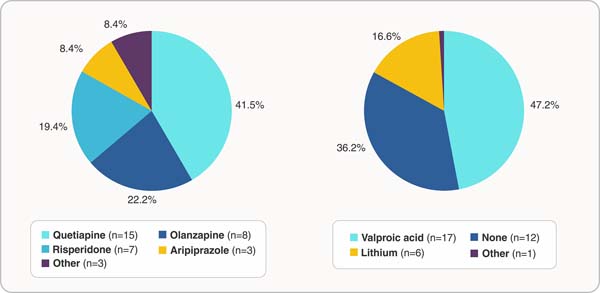
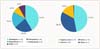
Quetiapine (41.6%) was the most commonly used antipsychotic after hospitalization, followed by olanzapine (22.2%) and risperidone (19.4%). Valproic acid the most commonly used mood stabilizer (47.2%). Lithium was used by 16.6%, and 33.3% had not started mood stabilizers (Figure 1).
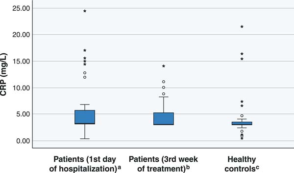
The BD group's serum C-reactive protein (CRP) levels on first day of hospitalization (mean: 3.12 mg/L, 5.62 mg/L [SD, 5.57] mg/L) were significantly higher than those of the control group (mean: 3.00 mg/L, 3.98 mg/L [SD, 4.23] mg/L). In the third week of treatment, there were no significant differences in CRP levels between the BD and control groups (Figure 2).
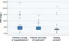
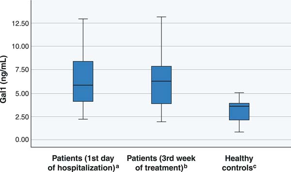
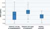
Gal-1 was significantly higher (mean: 6.11 ng/mL, 6.41 ng/mL [SD, 2.70] ng/mL) in the patient group on the first day of hospitalization than the control group (mean: 3.54 ng/mL, 3.20 ng/mL [SD, 1.09] ng/mL). Gal-1 levels did not differ significantly between the first day hospitalization and the third week of the treatment (Figure 3).
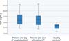
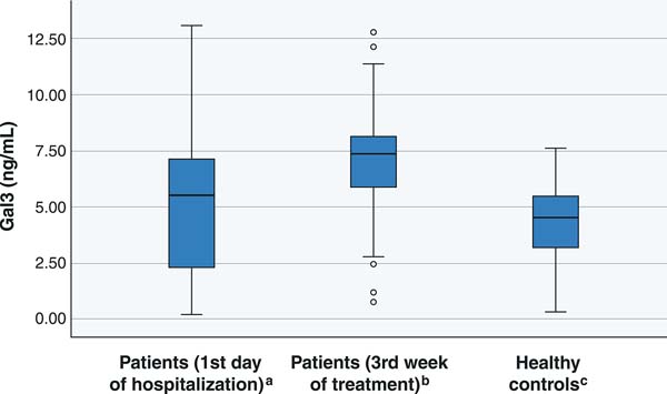
There were no significant differences in Gal-3 levels between patients on the first day of hospitalization and the control group. Patient Gal-3 levels in the third week of treatment (mean: 7.35 ng/mL, 6.88 ng/mL [SD, 2.77] ng/mL) were significantly higher than on the first day of hospitalization (mean: 5.56 ng/mL, 4.81 ng/mL [SD, 3.29] ng/mL) and those of healthy controls (p < 0.001) (Figure 4).
DISCUSSION
To the best of our knowledge, our study is the first attempt to assess the involvement of Gal in BD, including an examination of Gal levels before and after treatment. Our investigation yielded two notable findings concerning the etiopathogenesis of BD. First, Gal-1 levels were significantly higher in BD patients than healthy controls. Second, Gal-3 levels had increased significantly by the end of the third week of treatment. In addition, the finding that the CRP levels of patients were higher on the first day of hospitalization than in the third week of treatment and of healthy controls is important since it shows that inflammatory processes play a role in BD.
Previous studies have shown that immune system disorders that lead to neuroinflammation are involved in the pathophysiology of most chronic mental disorders.34 Current evidence supports the hypothesis that changes in the inflammatory system play a critical role in BD pathophysiology. Studies have shown that Gal plays an important role in immune and inflammatory response.27 Recent studies have proven the key role of Gal-1 and Gal-3 in neuroimmune response to various neuropathological conditions. In the light of these findings, we measured serum concentrations of CRP, Gal-1, and Gal-3 in BD patients either during a manic episode (acute exacerbation) or remission and in healthy controls, assessing the relationship between BD and inflammation, clarifying its possible role in the pathophysiology of the illness.
The fact that CRP levels were higher in patients than controls supports the hypothesis that inflammatory processes play a role in the etiopathogenesis of the illness. However, the fact that there was no difference in CRP levels between the acute and remission periods suggests that it is mostly related to chronic illness processes. According to Uyanık et al.,35 during manic episodes, patients with BD have significantly higher CRP levels before treatment, which decrease in the post-treatment phase compared to healthy controls. Our follow-up period was not long. Although CRP levels decreased between the manic episode and remission, this may be the reason why the difference was not significant.
Recent studies have shown that serum concentrations of Gal-1 change in neurological illness and increase during biological stress.36,37 Various studies have also shown that Gal-1 levels increase in cases of blood-brain barrier damage, oxidative stress, and neuroinflammation.36,38,39
Increased Gal-3 has been reported in various brain pathologies, including amyotrophic lateral sclerosis, Alzheimer's disease, and ischemic brain lesions. Dong et al.40 followed the functioning of 233 patients with acute ischemic attacks for 1 year, reporting that higher serum levels of Gal-3 were associated with lower disease severity and better functional recovery. However, Wang et al.41 observed higher Gal-3 levels in Alzheimer's patients and people with cognitive impairment than healthy controls, as well as a positive correlation between Gal-3 levels and Mini-Mental Status Examination scores.
In patients with schizophrenia, Kajitani et al.28 found equal Gal-1 levels to those of healthy controls but high Gal-3 levels. They also found that serum Gal-3 levels were positively correlated with positive symptom scores and negatively correlated with negative symptom scores. However, unlike other studies, Kılıç et al.30 reported that schizophrenia patients had lower Gal-3 levels than healthy controls and that there was a positive correlation between negative symptom scores and Gal-3 levels. Low Gal-3 levels may be due to neurodegeneration or to the patient group being less chronic. Yüksel et al.29 found a significant difference between serum Gal-1 levels of patients with schizophrenia, healthy siblings of schizophrenia patients, and a healthy control group, as well as that Gal-1 and Gal-3 levels were higher in the unaffected siblings of schizophrenia patients than the patients themselves or healthy controls. High concentrations of both Gal-1 and Gal-3 were detected in unaffected siblings, which suggests that Gal-1 and Gal-3 may protect against inflammation. Borovcanin et al.42 found that Gal-3 levels were lower during first-episode psychosis and relapse in schizophrenia patients than healthy controls, but they were higher in schizophrenia patients during remission.
Although different Gal-1 and Gal-3 levels were found in all these neuropsychiatric illnesses, these proteins are considered to play a neuroprotective role against increased neuroinflammatory response in most of them. Our results also support the neuroprotective role of Gal-1, given that patient Gal-1 levels were higher than controls. In light of these data, higher Gal-1 and Gal-3 levels were expected in the patient group than the control group, and the high Gal-1 values in the present study are consistent with the literature. If increased Gal-3 levels are a delayed reaction to increased inflammatory response, they should regress after the acute phase.42 The fact that Gal-3 was higher during remission than the acute phase in our patients suggests that it may play a role in adaptive late response to inflammation, as in schizophrenia. Methodological differences among studies may explain the inconsistent results. For example, CRP, Gal-1, and Gal-3 levels may have been affected by the number of patients included in the study, whether outpatients or inpatients were studied, antipsychotic use, and metabolic status (e.g., body mass index and cardiovascular risk factors).
One of this study's strengths is that the serum concentrations was re-evaluated after three weeks of treatment and compared with baseline. In a previous study of patients with schizophrenia, the only comparison was between the patient and control groups.28 In our study, the change in Gal levels after treatment was examined for the first time. Since no post-treatment evaluation had been previously undertaken, a 21-day period was determined in reference to a preclinical study.43 However, since the immunomodulatory response may differ in later periods, longer follow-up is required in future studies.
The present study has certain limitations. First, the findings cannot be generalized to larger populations due to the relatively small sample size, so studies with larger samples are needed. Second, we could not exclude the effects of antipsychotic medications on neuroinflammatory markers, but future studies could organize their analysis according to psychotropic drug groups. Third, we only assessed the early remission period of the illness. Re-assessment of serum concentrations during longer remission periods may provide a clearer picture of Gal's role in the pathophysiology of the illness. Finally, factors such as lifestyle, exercise, and nutritional changes can affect serum inflammatory processes. Although quite difficult, it would be useful to select patient groups with similar living standards in future studies.
In conclusion, it appears that Gal-1 may play a role in the pathophysiology of BD, while Gal-3 may serve as a candidate biomarker for treatment response. Our study is the first to show a change in Gal levels after BD treatment and to assess the role of Gal in BD. Further studies are needed with larger patient samples, longer follow-up, and neuroinflammatory response assessment according to psychotropic groups. Such research could contribute to our understanding of the etiopathogenesis of the disease and the development of potential new treatments, especially immunomodulatory ones.
ACKNOWLEDGEMENTS
The biochemical analyses in this study were supported by the Scientific Research Projects Unit of the Health Sciences University of the Republic of Turkey (Project No. 2021/150).
 PDF
 Print
 Send this article by email
 How to cite this article
 Submit a comment
 Mendeley
 Pocket
 Share on Facebook
 Share on Twitter