


| Note: How to cite this article: Sengül MCB, Agaçe M, Aygün D. Cerebellar atrophy associated with valproate-induced tremor: a case report based on three-dimensional volumetric magnetic resonance imaging. Braz J Psychiatry. 2026;48:e20254758. Epub 2026 Feb 16. http://doi.org/10.47626/1516-4446-2025-4758 |
| Received: December 11 2025; Accepted: January 20 2026 |
Psychotropic medications are essential to managing bipolar disorder, but their side effects can make it challenging for patients to adhere to treatment.1 Sodium valproate (VPA) is associated with elevated liver enzymes in almost 44% of patients, and it can also lead to tremors, hyperammonemia, and other metabolic disturbances.1 Most psychotropic medications do not cause structural brain changes that can be readily detected on imaging, as their effects typically occur at the molecular or cellular level. Even so, prolonged exposure to certain agents can occasionally lead to structural alterations that become visible in neuroimaging.2 VPA-induced cerebellar atrophy is a rare but serious adverse effect.3 Early recognition allows the drug to be discontinued promptly, supports both clinical and radiological recovery, and may prevent unnecessary investigations and consultations.4 This case report presents a patient with schizoaffective disorder and no prior neurological history who developed cognitive and neurological impairment during VPA treatment, accompanied by reversible cerebellar atrophy identified through three-dimensional brain volumetric (3D-BRAVO) magnetic resonance imaging (MRI). By utilizing 3D-BRAVO MRI for quantitative analysis, this report highlights the significant role of advanced volumetric imaging in objectively documenting the dynamic and reversible nature of VPA-induced cerebellar changes, thereby offering a more precise monitoring tool for clinicians compared to conventional qualitative imaging.
The patient, a 47-year-old single man living with his mother, provided written informed consent for case publication. He presented with symptoms including irritability, talking to himself, auditory hallucinations, suicidal thoughts, and reduced sleep, while inconsistently taking VPA 1000 mg/day, olanzapine 20 mg/day, quetiapine 800 mg/day, and chlorpromazine 50 mg/day. He had a 15-year history of schizoaffective disorder, including three previous hospitalizations: two for manic episodes and one for a depressive episode. On mental status examination, he exhibited disorganized behavior, irritability, and distractibility, along with auditory hallucinations and disorganized thought processes. He also showed persecutory and referential delusions, impaired judgment, lack of insight, psychomotor agitation, and reduced sleep.
The treatment was modified to VPA 1000 mg/day, olanzapine 20 mg/day, quetiapine 100 mg/day, and lurasidone 80 mg/day. The lurasidone dose was later increased to 160 mg/day. Despite these changes, his psychotic symptoms and agitation persisted, leading to the initiation of electroconvulsive therapy. After two ineffective sessions, the VPA dose was reduced to 500 mg/day (serum level of 12.5 mg/L while on 500 mg/day), chlorpromazine 100 mg/day was added, and the anesthetic agent was changed from propofol to ketamine. Additionally, a paliperidone long-acting injection (150 mg + 100 mg) was administered on day 8.
Two days after the eighth electroconvulsive therapy session, the patient presented with disorientation to time, place, and person, an inability to respond, and tremor (predominantly in both hands), with dysmetria and dysdiadochokinesia, prompting neurological evaluation. Electroencephalography revealed diffuse moderate abnormalities without lateralization or localization. Levetiracetam was administered intravenously at a dose of 18-20 mg/kg, but was then discontinued due to agitation. After a VPA load, as previously administered, elevated VPA levels were associated with worsening delirium and cerebellar signs. Extrapyramidal examination revealed rigidity in the right arm, and biperiden 4 mg/day was initiated. Laboratory tests for ongoing neurological symptoms were within normal limits.
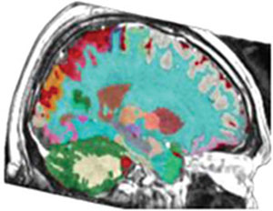
In 3D-BRAVO MRI, bilateral cerebellar hemisphere atrophy (more pronounced on the left side) was observed, along with an increased cerebrospinal fluid (CSF) space between the hemispheric sulci. Volumetric analysis using vol2Brain revealed CSF 117.03 cm3, cerebellum 112.24 cm3 (right 59.55 cm3, left 52.69 cm3), and cerebellar vermis 7.48 cm3 (Table 1).5 Given suspected VPA-related cerebellar atrophy, VPA was discontinued. Within two weeks, his tremor and cerebellar symptoms showed marked improvement, and his orientation returned to normal. At discharge, his treatment was revised to include olanzapine 10 mg/day, paliperidone long-acting injection 100 mg/month, and quetiapine 25 mg/day.
| Baseline (cm3) | Follow-up 8 months (cm3) | ||
 |
 |
||
| Structure | Baseline (cm3) | Follow-up 8 months (cm3) | Change (%) |
| Cerebrospinal fluid | 117.03 | 110.28 | -5.77 |
| Cerebellum - Right | 59.55 | 62.49 | +4.94 |
| Cerebellum - Left | 52.69 | 60.62 | +15.05 |
| Total cerebellum | 112.24 | 123.11 | +9.68 |
| Cerebellar vermis | 7.48 | 7.68 | +2.67 |
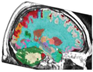
In 3D-BRAVO MRI, bilateral cerebellar hemisphere atrophy was observed, more prominently on the left side, with increased CSF space. Volumetric analysis using vol2Brain revealed increased cerebellar volumes and a reduction in CSF volume eight months after VPA was discontinued.

At the eight-month follow-up, 3D-BRAVO MRI revealed a CSF volume of 110.28 cm3, a total cerebellar volume of 123.11 cm3 (62.49 cm3 in the right hemisphere and 60.62 cm3 in the left), and a cerebellar vermis volume of 7.68 cm3 (Table 1). Compared with baseline, CSF volume decreased by 5.77%; right cerebellar hemisphere volume increased by 4.94%; the left hemisphere by 15.05%; total cerebellar volume by 9.68%; and the vermis by 2.67%. Psychiatric evaluation indicated a euthymic mood with no psychotic features, and the neurological examination revealed no residual cerebellar deficits.
Psychotropic medications offer important therapeutic benefits, yet they can also lead to adverse effects. VPA is a widely used treatment for bipolar and schizoaffective disorders and is generally well tolerated.2 However, VPA can also lead to serious complications, including hyperammonemia and fulminant hepatic encephalopathy. More recently, a fourth form of VPA-related encephalopathy, referred to as "reversible cerebral and cerebellar pseudoatrophy," has been identified.4 The earliest reported cases date to 1987, when McLachlan observed reversible brain atrophy with computed tomography during VPA monotherapy.3
In adults, cerebral and cerebellar pseudoatrophy could manifest through a variety of neurological and cognitive symptoms, including ataxia, parkinsonism, tremor, dysmetria, dysdiadochokinesia, cognitive slowing, stupor, bradykinesia, memory impairment, amimia, inappropriate smiling, hallucinations, and disorientation.4 In our patient, 12 years of VPA exposure was associated with tremor, disorientation, and cerebellar signs consistent with cerebellar atrophy, despite normal laboratory findings. Notably, his symptoms improved within two weeks of discontinuing VPA, in line with previous reports.1-4 Pediatric reports indicate that imaging abnormalities may resolve within 4 months to 2 years after discontinuing VPA, although the product information reports a potential for irreversible cerebral and cerebellar atrophy.4
The mechanism underlying VPA-related cerebellar effects remains unclear, although hyperammonemia has been proposed as a contributing factor.3,6 Notably, CSF ammonia and glutamine levels may be elevated even when serum ammonia remains within normal limits.7 In our patient, liver function tests and serum ammonia were normal, VPA serum levels were low, and CSF analysis was not performed. Symptoms can emerge either soon after treatment begins or following prolonged exposure.8 Although cerebellar volume increased over time, it remains uncertain whether the observed atrophy represented a lasting change or a reversible process. Consistent with much of the literature, our report is limited by a lack of baseline neuroimaging prior to initiating VPA treatment. While this precludes a direct comparison with the patient's pre-treatment cerebellar volume, the objective increase in volume following VPA discontinuation strongly supports a drug-induced etiology.
If neurological symptoms or imaging changes indicate VPA-related cerebellar atrophy, VPA should be stopped promptly, regardless of treatment duration or serum VPA level. Early withdrawal can help prevent unnecessary investigations or interventions, and most patients begin to show meaningful clinical improvement within 2-3 weeks.8
 PDF
 Print
 Send this article by email
 How to cite this article
 Submit a comment
 Mendeley
 Pocket
 Share on Facebook
 Share on Twitter