The hippocampus has been a focus of attention in neuroimaging studies of bipolar disorder (BD). This structure has been recognized as particularly vulnerable to the neurotoxic effects of obesity, and it is apparently subject to synergistic, deleterious action from obesity and mood disorders.
1 Comorbid obesity is highly prevalent in people with BD.
2,3 In addition, there is a robust body of evidence showing that obese individuals with BD have poor cognitive performance and increased levels of inflammatory mediators as compared to those with normal weight.
4 Thus, the objective of this study was to ascertain whether an association exists between body mass index (BMI) and hippocampal volume in individuals with BD and to compare hippocampal volumes between patients and healthy controls.
Sixty-five subjects were enrolled: 26 patients with BD in euthymia and 39 controls. Inclusion criteria for patients were: a) age > 18 years; b) DSM-IV criteria for bipolar I; and c) criteria for remission, defined as scores < 7 on the 17-item Hamilton Depression Scale (HAM-D) and on the Young Mania Rating Scale (YMRS) for at least 1 month prior to assessment. Patients with severe clinical illnesses were excluded. The control group consisted of healthy volunteers who had no current or previous history and no first-degree family history of a major psychiatric disorder, including dementia, as assessed by the non-patient version of the Structured Clinical Interview for DSM-IV. Written informed consent was obtained from all subjects in accordance with the Declaration of Helsinki, and the study protocol was approved by the local ethics committee.
Magnetic resonance imaging data were obtained in a Philips Achieva 1.5-tesla scanner (Amsterdam, the Netherlands). T1 high-resolution sagittal 3D magnetization-prepared rapid acquisition gradient-echo (MPRAGE) sequences were acquired with NEX = 1, image matrix = 256 x 232, flip angle = 8 degrees, echo time = 4 ms, repetition time = 8.63 ms, and voxel size 1 x 1 x 1 mm
3, yielding 160 slices. Subcortical volumetric segmentations were performed with the Freesurfer image analysis suite, version 5.1.0 (
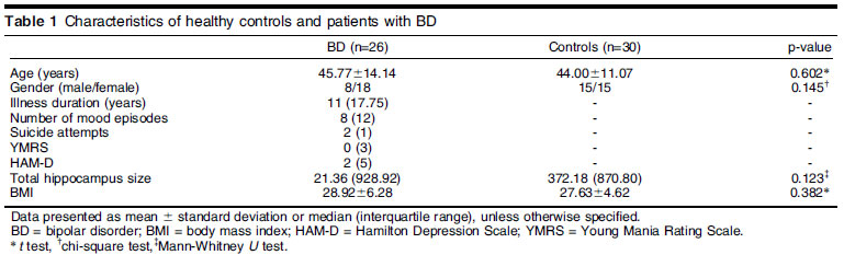
http://surfer.nmr.mgh.harvard.edu/). Intracranial volume was regressed out from hippocampal volume. Descriptive analyses are presented as mean (standard deviation) or median (interquartile range). Appropriate tests for parametric or nonparametric distribution are indicated in Table 1. Correlations were obtained by Spearman rank correlation coefficients.
Clinical and demographic characteristics of the sample are summarized in Table 1. There was no significant difference in total hippocampus size between patients and controls (p = 0.123). There was no correlation between total hippocampus size and BMI in the overall sample (p = 0.153, rho = -0.194), nor separately in the BD (p = 0.084, rho = -0.345) and control (p = 0.823, rho = -0.043) groups. Significant BMI and BD diagnosis interactions for white and gray matter reductions have been described
3,5; furthermore, obesity in BD has been associated with worse disease progression, suggesting that nutritional interventions are highly desirable for better outcomes. Although obesity is a potentially modifiable condition that is highly prevalent in individuals with BD and the hippocampus is very susceptible to the neurotoxic effects of obesity, this study did not find an association between hippocampus size and BMI.
DISCLOSUREThe authors report no conflicts of interest.
REFERENCES1 Fotuhi M, Do D, Jack C. Modifiable factors that alter the size of the hippocampus with ageing. Nat Rev Neurol. 2012;8:189-202.
2 Bond DJ, Ha TH, Lang DJ, Su W, Torres IJ, Honer WG, et al. Body mass index-related regional gray and white matter volume reductions in first-episode mania patients. Biol Psychiatry. 2014;76:138-45.
3 Bond DJ, Kauer-Sant'Anna M, Lam RW, Yatham LN. Weight gain, obesity, and metabolic indices following a first manic episode: prospective 12-month data from the Systematic Treatment Optimization Program for Early Mania (STOP-EM). J Affect. Disord. 2010;124:108-17.
4 Yim CY, Soczynska JK, Kennedy CH, Woldeyohannes HO, Brietzke E, McIntyre RS. The effect of overweight/obesity on cognitive function in euthymic individuals with bipolar disorder. Eur Psychiatry. 2010;27:223-8.
5 Bond DJ, Lang DJ, Noronha MM, Kunz M, Torres IJ, Su W, et al. The association of elevated body mass index with reduced brain volumes in first-episode mania. Biol Psychiatry. 2011;70:381-7.





 © 2026 All rights reserved


 PDF
 Print
 Send this article by email
 How to cite this article
 Submit a comment
 Mendeley
 Pocket
 Share on Facebook
 Share on Twitter