


ABSTRACT:
OBJECTIVE: Panic disorder (PD) is a common disabling condition characterized by recurrent panic
attacks. Emotional and behavioral impairments are associated with functional connectivity
(FC) and network abnormalities. We used whole-brain FC, modular networks, and graph-theory
analysis to investigate extensive network profiles in PD.
METHODS: Functional magnetic resonance imaging (fMRI) data from 82 subjects with PD and 97
healthy controls were included. Intrinsic FC between each pair of 160 regions, six
intra-network, and 15 inter-network FCs were analyzed. Topological properties were
explored.
RESULTS: PD patients showed altered FCs within the right insula, between frontal cortex-posterior
cingulate cortex (PCC), frontal cortex-cerebellum, and PCC-occipital cortex (corrected
p < 0.001). Lower connections within the sensorimotor network (SMN) and SMN-occipital
network (OCN) were detected (p < 0.05). Various decreased global and local network
features were found in PD (p < 0.05). In addition, significant correlations were found
between PD symptoms and nodal efficiency (Ne) in the insula (r = -0.273, p = 0.016) and intra-insula FC (r = -0.226, p = 0.041).
CONCLUSION: PD patients present with abnormal functional brain networks, especially decreased
FC and Ne within the insula, suggesting that dysfunction of information integration
plays an important role in PD.
Keywords: Panic disorder; functional connection; modular network; topology properties; insular cortex.
FIGURES
| Received: December 20 2023; Accepted: March 03 2024 |
INTRODUCTION
Panic disorder (PD) is one of the most common anxiety conditions, with lifetime prevalence ranging from 1 to 5%.1 It is characterized by the sudden onset of intense fear, physical discomfort, a feeling of impending doom or death, loss of control, anticipatory anxiety, and avoidance. Because it is typically chronic and prone to recurrence, PD causes prolonged impairment of social function and mental disabilities,2 and is associated with somatic comorbidity, such as stroke, further complicating the patient's overall health status and treatment outcomes.3 PD symptom profiles are linked to local and network abnormalities.4 Altered functional connectivity (FC), e.g., limbic areas and connection with sensory and motor regions,5 the anterior cingulate cortex and the precuneus, has been found in PD patients.6 Using voxel-wise independent component analysis, Ni et al.7 showed decreased FC in the sensorimotor network (SMN), default mode network (DMN), and cerebellar network in PD. However, detailed network abnormalities and interactions in PD remain largely unclear.
The critical clinical manifestations of PD, such as abnormal body sensations,8 highlight the role of insular cortex hyperactivation in autonomic regulation.9 In addition, its connection with various brain regions also impacts the gastrointestinal tract and heart,10 contributing to abundant somatic symptoms. For instance, increased FC between the insula and thalamus has been reported in rest-state functional magnetic resonance imaging (fMRI).11 It was assumed that the insula was altered and actively affected brain networks in PD; however, the exact mechanism was unclear.
The insula is a crucial region in the SMN12 and the salience network (SN).13 These networks participate in sensory processing, motor, stimulus, internal environment monitoring, and other functional domains. When the SMN is processing sensory and motor information, the SN will help select and pay attention to the information currently crucial to the individual and ensure the allocation of attention resources at the right moment and under the right conditions.14 Notably, the insula is an SMN and SN connecting node, contributing to stimulus-driven processing and sustaining a stable internal environment.15
However, these network interactions need to be investigated on a larger map, considering the potential effect of other networks. A disbalance between SMN and the default network has been observed in bipolar disorder.16 In contrast, complex relationships between the default network, central executive network, SN, and sensory network have been detected in schizophrenia, suggesting that different psychopathological states may be underpinned by distinct patterns of functional organization in the brain.17 A scaled correlation analysis showed increased FC between the somatosensory cortex and the thalamus in PD.18 Lai et al.19 revealed the importance of the connection between limbic areas and sensory and motor regions using network-based statistics of the connectome in PD patients. These SMN-centered results are quite different from the findings of FC alterations in other anxiety disorders, such as social anxiety and posttraumatic stress disorder,20 underscoring the unique impact of PD on the brain's functional organization. The alterations in the SMN seen in PD patients highlight a crucial role for this network.
The altered network topology attribution of PD needs to be understood. Notably, the topology reveals the interaction and communication patterns between brain nodes, affecting emotion and behavior.21 Disrupted network topology properties are associated with severe brain disorders, including Alzheimer's disease22 and schizophrenia,23 as well as with treatment response.14 Studies have shown that, in social anxiety disorders, decreased nodal local efficiency (NLe) in the left posterior cingulate cortex (PCC) was significantly associated with avoidance and anxiety-related symptoms.24 However, little is known about how network attributes are altered in patients with PD.4
The aim of this study was to investigate the status of FC networks (such as insular cortex) and modular network (such as SMN) interactions. In addition, we analyzed the topological properties of brain networks. Finally, we explored their relationships with the clinical symptoms of PD. Our hypotheses were as follows: 1) the connectome, limbic, and insula connections, in particular, would be decreased in the PD patients; 2) the PD group would exhibit a significant decrease in FC of networks correlated with SMN, and this would be the core network of intra-connectivity alterations; and 3) insular nodal efficiency (Ne) would be significantly reduced in PD patients, which may be a crucial factor contributing to disrupted global information transmission and integration, ultimately affecting the clinical symptoms of PD as operationalized by measures such as the Panic Disorder Severity Scale (PDSS).
MATERIALS
Participants
Participants with PD were recruited from the Department of Psychosomatics and Psychiatry, Zhongda Hospital affiliated with Southeast University, from March 2020 to December 2022. Two senior psychiatrists reached a consensus on PD diagnoses according to the DSM-5. The detailed inclusion criteria were as follows: 1) inpatients; 2) age 18 to 65 years; 3) right-handedness; 4) no severe or unstable organic brain diseases or somatic conditions (cardiac, hepatic, renal, endocrine, digestive, hematological, etc.); 5) normal laboratory and ancillary test results; 6) absence of other major psychiatric disorders, including personality disorders and substance-related disorders; 7) no primary neurological disorders, such as dementia or stroke; and 8) no significant structural abnormalities on T1-weight images and no major white-matter changes such as infarction or other vascular lesions on T2-weighted MRI. PD participants underwent both clinical assessment and MRI at baseline, followed by an additional clinical assessment without MRI 2 weeks later.
Healthy controls (HCs), aged 18 to 65, were recruited from the local community through advertising in Nanjing. Eligibility criteria for HCs included no mental disorders, Hamilton Anxiety Rating Scale (HAMA) < 8, and 17-item Hamilton Depression Rating Scale (HAMD-17) < 8, as assessed by two psychiatrists using the Structured Clinical Interview for DSM, fourth edition (DSM-IV) Axis I Disorders (SCID-I/P). Psychiatric or severe organic illness were exclusion criteria. After a quality check, data were available for 82 participants with PD (30 males and 52 females) and 97 HCs (35 males and 62 females).
Assessment tools
The PDSS and Panic-Associated Symptom Scale (PASS) served as the primary tools to evaluate panic symptoms in PD patients at baseline. The HAMA and HAMD-17 were used to assess anxiety and depression symptoms, respectively. Higher scores indicate more severe symptoms. These patients were under that medication prescribed by their psychiatrists, medications status was as follows: 72 patients received selective serotonin reuptake inhibitors (SSRIs), of which 46 were escitalopram, 19 were paroxetine, and the others seven included fluoxetine and sertraline; 10 patients with serotonin-norepinephrine reuptake inhibitors, of which seven were venlafaxine, and three were duloxetine. In this study, the doses of antidepressants were all converted to fluoxetine equivalents.25 The conversion method used is as follows: 20 mg/d for fluoxetine is respectively equivalent to 9 mg/d for escitalopram, 17 mg/d for paroxetine, 49.3 mg/d for sertraline, 74.7 mg/d for venlafaxine, and 60 mg/d for duloxetine.
MRI data acquisition, pre-processing
All participants underwent the MRI scans in a 3.0-Tesla superconducting MRI system (Siemens Medical Systems, Erlangen, Germany) at Zhongda Hospital. PD participants were scanned within 7 days of hospitalization. The scan was scheduled to occur within the time frame of 12:00-2:00 p.m. to minimize the effects of diurnal variations. Participants lay supine with the head snugly contained by special head pads to reduce head motion, and they wore headphones to minimize the effect of machine noise during the scan. All participants were instructed to keep their eyes closed, remain awake, relax, and not think about anything specific during the scan. Three-dimensional T1-weighted images were recorded using magnetization prepared rapid gradient echo (MP-RAGE) sequences: repetition time (TR) = 1,900 ms, echo time (TE) = 2.48 ms, flip angle (FA) = 9°, acquisition matrix = 256 × 256, field of view (FOV) = 250 × 250 mm2, thickness = 1.0 mm, gap = 0, slices = 176, time = 4 min 18 s. The acquisition parameters of resting-state fMRI were used by a gradient-recalled echo-planar imaging pulse sequence with the following parameters: TR = 2,000 ms, TE = 25 ms, FA = 90°, acquisition matrix = 64 × 64, FOV = 240 × 240 mm2, thickness = 3.0 mm, gap = 0 mm, 32 axial slices, 240 volumes, 3.75 × 3.75 mm2 in-plane resolution parallel to the anterior-posterior commissure line, time = 8 min.
The images were preprocessed using the Data Processing Assistant for Resting-State Function (DPARSF 5.2 Advanced Edition) MRI toolkit, which integrates procedures based on the Resting-State fMRI Toolkit (REST) (http://www.restfmri.net) and statistical parametric mapping software package (SPM12) (http://www.fil.ion.ucl.ac.uk/spm). The specific steps were processed sequentially as follows: 1) convert the DICOM file to NIFTI format data; 2) remove the first 10 time points to exclude data instability due to inherent scanner noise; 3) slice timing and correct for head motion (participants with more than 2 mm or 2° of motion were excluded from the study); 4) co-register T1 to functional image and reorient; 5) normalize T1-weighted anatomic images to the Montreal Neurological Institute space; 6) perform spatial smoothing (6 mm); 7) remove the linear trend within each voxel's time series; 8) regress out nuisance signals (white matter, cerebrospinal fluid signals and global signal); and 9) apply a temporal bandpass filter (0.01-0.08Hz).
Generation of functional connectomes
To generate whole-brain functional connectomes, we parcellated each participant's brain image into 160 brain regions or nodes sufficiently covering most of the cortical areas and the cerebellum based on the Dosenbach atlas26 (5-mm spheres for each region of interest [ROI]). This atlas has been widely used to analyze whole-brain FC of resting-state networks. We computed the correlation matrices by extracting the time course of each ROI. The Pearson correlation coefficient (r) was calculated between the time course of one ROI and all other ROIs. Then, 160 × 160 correlation matrices based on the preprocessed data above were obtained by static correlation calculation using Gretna software (https://www.nitrc.org/projects/gretna/). The 160 brain regions were divided into six networks according to the Dosenbach atlas: DMN, fronto-parietal network (FPN), cingulo-opercula network (CON), SMN, occipital network (OCN), and cerebellum network. These networks are respectively labeled as 1-6 (Supplementary Table S1). Analysis of intra- and inter-network FC was performed by Gretna software. Intra-network connectivity refers to the FC within the same neural network, indicating the level of synchronization or communication among brain regions that are part of the same functional network. Inter-network connectivity, on the other hand, pertains to the functional connections between different neural networks, highlighting how these distinct networks communicate and coordinate with each other. Subsequently, every comparison measurement between two networks or within each network was converted to area under the curve (AUC) values by a MATLAB (R2022a) script to provide a scalar that did not depend on specific threshold selection for further analysis. The results of altered FC in PD patients were visualized in BrainNet Viewer.
Analysis of network topology
Graph theory has played an important role in characterizing topological organization and making inferences about the flow of information through brain networks.21 The topological characteristics of global and local brain network nodes were measured with 0.01 steps in the sparsity range of 0.039 to 0.4 using Gretna software, and the Dosenbach 160 brain atlas was used as a template.
Characteristics included global efficiency (Eg), local efficiency (Eloc), and small-worldness. The index of the small-world network includes the clustering coefficient (Cp), characteristic path length (Lp), small-worldness (sigma), normalized Cp (gamma), and normalized characteristic Lp (lambda). Eg quantifies a network's capacity for information dissemination, determined by the mean of the reciprocal values of the shortest Lps connecting every node within the network. An elevated Eg signifies rapid dissemination of information and a closely-knit topological arrangement among the network's nodes. The Eloc of a network measures the communication efficiency among the neighbors of a given node when that node is removed. Cp gauges a network's local interconnectivity based on the average Cp across all nodes, reflecting the density of interconnections within localized regions. Lp, representing the mean shortest distance (counted as the smallest edge count) that connects any two nodes, serves as a measure of the network's overall navigability. Characterized by a high Cp and a low Lp, a small-world network optimizes both localized interconnectivity and wide-ranging network reach. Small-worldness is quantitatively assessed through comparison of the Cp and Lp of the actual brain network against those of a hypothetical random network.21
Local characteristics included Ne, NLe, nodal Cp (NCp), nodal shortest length (NLp), and nodal degree centrality (NDc). The result of altered local properties in PD patients was visualized in BrainNet Viewer. Ne at a nodal level assesses how effectively a node facilitates information flow to other nodes across the network, characterizing the efficiency of parallel information transmission by the node within the network. NLe measures the communication efficiency among its neighboring nodes when that node is removed. NCp, indicating the proportion of actual to potential connections among a node's neighbors, reflects the node's role in fostering a tightly knit, resilient network structure capable of withstanding disruptions. NLp quantifies the average distance or routing efficiency between that node and all other nodes within the network. Finally, NDc is determined by its total edge count, indicating its relative importance within the network. A node with higher degree centrality is deemed crucial for the network's functionality.21
Statistical analysis
The data was analyzed using SPSS version 25. All data used for parametric testing were confirmed to conform to a normal distribution, as determined by the Kolmogorov-Smirnov (K-S) test, with p-values greater than 0.05. An independent two-sample t test and chi-square test were applied to determine significant differences in demographic data and assessment scores between PD and HCs. Continuous variables were presented as mean ± SD. Categorical variables were presented as numbers or percentages.
The FC differences between the PD group and HCs were analyzed by two-sample t-tests with false discovery rate (FDR) correction. Two-sample t-test analysis of each two networks or within each network AUC was used to calculate intra- and inter-network FC differences between the two groups. Similarly, AUC values were extracted for network attributes to further analyze two-sample t-tests and correlations.
The relationships between FC and network attributes and clinical features were revealed by bias correlation analysis using age, sex, head motion, and medications status as covariates in the PD group. The threshold of statistically significant differences was defined as p < 0.05.
Ethics statement
The study adhered to the Declaration of Helsinki and received approval from the ethics committee of Zhongda Hospital affiliated with Southeast University (approval no. 2022ZDSYLL194-P01). Participants provided written informed consent.
RESULTS
Demographic and clinical data
A total of 179 participants, 82 PD patients (30 males and 52 females) and 97 HCs (35 males and 62 females), were included in this study. There were no significant differences in terms of age, sex, and body mass index (BMI) between the two groups, but the PD group had fewer years of education than the HCs. At baseline, HAMA and HAMD-17 scores differed significantly between groups. PDSS and PASS scores showed a marked decrease following a 2-week treatment period in PD patients. A detailed comparison of demographic and clinical data between the two groups is provided in Table 1.
| PD (n=82) | HCs (n=97) | t/?2 | p-value | |
|---|---|---|---|---|
| Age, years | 40.67±12.40 | 37.65±12.64 | 1.607 | 0.110┼ |
| Sex, male/female | 30/52 | 35/62 | 0.005 | 0.944╬ |
| BMI (kg/m2) | 23.25±3.99 | 23.23±3.10 | 0.029 | 0.977┼ |
| Education, years | 11.93±4.06 | 15.57±4.35 | -5.740 | < 0.001* ┼ |
| Duration of illness, months | 22.48±38.71 | - | - | - |
| PDSS at baseline | 13.09±3.80 | - | - | - |
| PASS at baseline | 11.22±3.71 | - | - | - |
| HAMA at baseline | 21.67±6.39 | 1.81±1.85 | 29.193 | < 0.001* ┼ |
| HAMD at baseline | 13.01±5.46 | 1.32±1.66 | 20.016 | < 0.001* ┼ |
| PDSS at week 2 | 4.35±3.49 | - | - | - |
| PASS at week 2 | 3.12±2.51 | - | - | |
| HAMA at week 2 | 6.65±3.80 | - | - | - |
| HAMD at week 2 | 3.71±2.02 | - | - | - |

Analysis of differences in whole-brain functional connectivity between the two groups
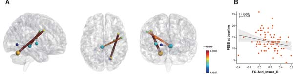
Analysis of FC between brain regions revealed four edges across seven nodes that differed between groups when controlling for confounders (including sex, age, and head motion) and applying FDR correction. These included connections between the right mid-insula (across various networks), right occipital and left PCC, right frontal cortex and left PCC, and right lateral cerebellum and dorsal frontal cortex (Figure 1A and Supplementary Table S2).
Further correlation analysis showed that right mid-insula FC was negatively correlated with PDSS scores (Figure 1B). Additionally, we differentiated males and females in the aforementioned correlation plots using distinct colors (Supplementary Figure S1).

Comparison of brain network changes between subjects with panic disorder and health controls
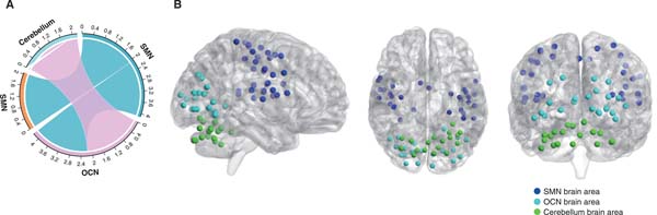
Analysis of intra-network and inter-network FC was conducted for the two groups after controlling for age, sex, and head motion. PD patients exhibited decreased FC in SMN-SMN, SMN-OCN, and OCN-cerebellum (Figure 2 and Supplementary Table S3). No significant differences were observed in other intra-network or inter-network FC measures.

Difference analysis of brain topology between the panic disorder and healthy control groups
Global topology property differences
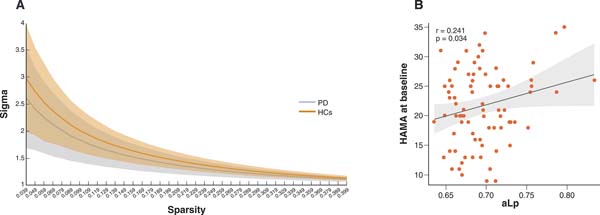
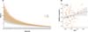
Both PD patients and HCs displayed characteristics consistent with small-world networks (sigma > 1) across varying sparsity levels (Figure 3A). The PD group showed significantly lower AUC values of sigma, gamma, and Eg compared with the HC group. Conversely, an increased AUC value for Lp was observed in PD patients. The two groups had no significant differences in other global properties (Table 2). A positive correlation was noted between AUC of characteristic path length (aLp) and HAMA scores at baseline in the PD group (Figure 3B).
| Global property | PD | HCs | t | p |
|---|---|---|---|---|
| aEg | 0.199±0.008 | 0.201±0.006 | -2.150 | 0.033* |
| aEloc | 0.240±0.016 | 0.243±0.014 | -0.960 | 0.338 |
| aCp | 0.164±0.026 | 0.164±0.022 | 0.148 | 0.883 |
| aLp | 0.694±0.037 | 0.682±0.033 | 2.285 | 0.023* |
| aSigma | 0.527±0.08 | 0.564±0.077 | -3.159 | 0.002* |
| aGamma | 0.553±0.088 | 0.595±0.084 | -3.249 | 0.001** |
| aLambda | 0.373±0.004 | 0.374±0.005 | -1.208 | 0.229 |
Sigma, small-worldness. Negative t-values denote the global property was lower in the PD group than in the HC group; positive t-values denote it was higher in the PD group.
aCp = AUC of clustering coefficient; aEg = AUC of global efficiency; aEloc = AUC of local efficiency; aGamma = AUC of normalized clustering coefficient; aLambda = AUC of normalized characteristic path length; aLp = AUC of characteristic path length; aSigma = AUC of small-worldness; HCs = healthy controls; PD = panic disorder.
* p < 0.05, ** p < 0.01.

Nodal property differences
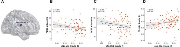
Compared with the HC group, the AUC value of Ne was significantly decreased in the right middle insula in the PD group (Figure 4A) (t = -3.882, p < 0.001 by FDR correction), which is located in area 103 of the Dosenbach 160 atlas. No significant difference was found in AUC of NCp, AUC of NLp, AUC of NLe, or AUC of NDc between the two groups (all controlled for confounders, including sex, age, and education, and corrected by FDR).

Furthermore, negative correlations were found between AUC of nodal efficiency (aNe) of the right middle insula and PDSS/PASS scores at baseline (Figure 4B and 4C). Again, we differentiated males and females in the aforementioned correlation plots using distinct colors (Supplementary Figure S2).
The differential brain area was all in the right middle insula in aNe, and further correlation analysis showed that aNe of Mid_Insula was positively correlated with FC between Mid_Insula_R (Figure 4D). Then we also used different dot colors to differentiate men vs. women (Supplementary Figure S3).
DISCUSSION
Our study has three main findings: firstly, PD revealed various abnormalities in connectivity, including in the frontolimbic (insula and frontal cortex in particular), sensory, and perceptual processing regions, as well as in the cerebellum. Secondly, patients with PD showed lower connectivity within the SMN, SMN, OCN, and OCN-cerebellum than HCs. Thirdly, patients with PD exhibited deficits of Ne in the right insula and abnormal global features. In addition, aberrant connectivity and topological attributes correlated with the severity of anxiety and PD-related symptoms.
PD features autonomic system responses, such as rapid heart rate and shortness of breath. The insula has a wide range of functions in the human autonomic system, ranging from sensorimotor and emotional processing to higher-level cognition. Studies have indicated an important role of the insula in cardiovascular regulation and pulmonary sensation.10 According to associated studies about functions and connections, multimodal sensory information including visual, auditory, gustatory, olfactory, and tactile inputs may converge into the insular cortex.10 The insula, which integrates the information entered into the thalamus by sensory organs,27 is a vital input center of visceral sensation, and abnormalities within it will arouse aberrant interoception (i.e., the sensation of the physiological state of the body).28 Furthermore, the insula and cingulate brain regions are considered integral parts of a "fear-conditioning" circuitry, and the SN that drives interoception in particular.29 Our finding reveals that intra-insula connectivity is lower in patients with PD, partly in line with previous research.9 Simultaneously, we showed that PDSS scores increase as intra-insula FC increases. Abnormality in the insula-insula loop may be an important neural correlate of PD. Decreased intra-insula FC appears to predict dysfunction of information integration. In addition to its role in processing sensory information, the insula also plays a crucial role in threat perception and the integration of motor information, such as avoidance behavior.10,28 This connection stems from the insula's capacity to amalgamate somatosensory, autonomic, and emotional data to steer behaviors in situations that may pose a threat.
Functionally synergistic and integrated networks underlying cognitive and emotional processes are the essence of the brain.30 The brain's modular network organization, characterized by tightly connected clusters that facilitate quick and efficient information flow and integration, plays a crucial role.21 The insula, part of the SMN, is involved in sensory information processing and motor functions.31 The occipital cortex, crucial for visual information processing and perception of facial affect and environmental change, communicates extensively with the cerebral cortex.32 Kim et al.20 summarized that hyperconnectivity in SMN might arouse abnormally high sensitivity of interoception and somatosensory stimuli, underlying the typical somatic symptoms of PD. However, the present study showed that the FC of intra-SMN and SMN-OCN was decreased in patients with PD, which suggested low communication efficiency and dysfunction of intra- and inter-module communication. Simultaneously, this finding enhanced the potential background of the SMN for panic attack-related somatosensory symptoms (including paresthesia, heat or cold sensation, and higher skin conductance) and motor abnormalities (e.g., fright, fear of losing control, and phobic avoidance), which are usually observed in PD.8,19 Our results differ from those of the previous study, perhaps due to the use of different methods and brain atlases. To summarize, emerging evidence consistently reveals that abnormalities of the SMN in PD appear to play a prominent role in interoception regulation and motor function, and the SMN might be a marker of vulnerable to panic attacks.20
Graph theory is a powerful tool for characterization of regional and global topological signatures of brain functional networks. At the local level, our findings in this study showed decreased Ne of the insula in PD patients. Ne represents the parallel information transmission efficiency of a brain node.4 The insula, densely connected within the brain hemisphere,10 also has direct structural connections to the autonomic nervous system.28 Decreased Ne in the insula region revealed reduced communication efficiency from the perspective of network information-carrying, and the negative association found between aNe of the insula and PDSS/PASS scores indicates that decreased transmission efficiency of insula exacerbates panic symptoms in PD patients. Notably, intra-insula FC correlated negatively with aNe of the insula, which indicates that information integration dysfunction may affect the efficiency of brain network information transmission in PD.
Small-world networks, characterized by high Cp and low characteristic Lp, facilitate rapid information transmission. At the global level, our study found that PD patients and HCs both exhibited small-world attributes (sigma > 1), with PD patients showing decreased small-worldness. This decrease might relate to impaired remote communication across different brain regions in PD. Our findings also support previous reports that observed similarly reduced small-worldness in PD4 and social anxiety disorder.33 Decreased Eg, normalized Cps, and increased Lp in PD patients, suggest disrupted brain networks and global information transmission from the perspective of functional integration and separation in PD. Only a lower Lp assures the transmission capacity of global information and supports a reliable basis of functional integration of the brain. Furthermore, correlations between HAMA scores and Lp in PD patients indicate that disruptions in global information transmission may worsen anxiety symptoms.
Moreover, our study found a positive correlation between the PCC and the right frontal and occipital cortex. The PCC is the hindmost portion of the cingulate cortex,34 involved in a wide range of attentional processes: self-monitoring, remembering the past, thinking about the future, and assessing the environment.35 The frontal cortex plays a significant role in cognitive processing36 and processing higher-order sensory information,37 and the symptoms of PD were related to incorrect input of signals to the frontal and limbic regions. Sensory information hypersensitivity was theoretically suppressed by the frontal cortex,37 and this suppressed pattern appeared useless in the long run. Patients with PD may overuse maladaptive regulatory strategies,38 become exhausted, and even arouse more hypersensitive anticipatory anxiety.39 Moreover, increased posterior cingulate and occipital cortex connectivity may indicate a perceptual visual bias, which could lead to emotional and somatic symptoms.
Additionally, some evidence indicates that, besides controlling movement, the cerebellum was also engaged in emotion regulation, attention, cognition, and fear conditioning consolidation, which might correlate with PD.7 The cerebellum also controls somatosensory processing.7 Therefore, the increased FC between the dorsal frontal cortex and cerebellum and the decreased FC of OCN-cerebellum observed in the current study might be associated with affectional and cognitive processing dysfunctions in PD.
It is worth noting that sex could influence these alterations in brain function. Ohrmann et al.40 found that, when responding to angry facial expressions, women demonstrated significantly stronger connectivity between the amygdala, prefrontal cortex, and thalamus compared to men. These results emphasize sex as an important variable in brain connectivity. Moreover, age-related changes in brain function can alter neurocircuitry,36 potentially influencing susceptibility to, and the manifestation of, neurological and psychiatric conditions. However, no studies have yet reported on the relationship between age and neurocircuitry in PD. Future research should specifically aim to dissect the complex interactions between age, sex, and brain neurocircuitry changes. Such studies would greatly enhance our understanding of the underlying mechanisms and contribute to the development of more personalized therapeutic approaches.
In this study, we conducted another round of clinical psychological assessments 2 weeks after enrollment. Regrettably, we did not find a relationship between 2-week efficacy (represented by the PDSS reduction rate) and FC, modular networks, or topological structure. However, PD is characterized by a chronic course, making the durability of treatment outcomes crucial.41 Thus, if the duration of treatment is extended, outcomes may be potentially affected. Additionally, in future follow-up MRI studies, the FC and brain networks that changed from baseline might return to normal. This will be a key focus of our research moving forward.
There are several limitations to our study. First, we used the Dosenbach 160 functional atlas; future studies need to consider other atlases as well. Second, medication use is inevitable in PD and might affect functional imaging findings. Third, follow-up scans were not collected in this study. Longitudinal research might help elucidate how the brain networks change over time in PD.
In conclusion, we found aberrant connectivity profiles and local properties in the insula and related networks, indicating their essential roles in information integration, somatosensory symptoms, and escape/avoidance behavior in PD. These findings provide new information and perspectives for PD.
ACKNOWLEDGEMENTS
This work was supported by the Postgraduate Research & Practice Innovation Program of Jiangsu Province (SJCX22_0071).
The authors thank the participants who generously gave their time to this study.
 PDF
 Print
 Send this article by email
 How to cite this article
 Submit a comment
 Mendeley
 Pocket
 Share on Facebook
 Share on Twitter