


ABSTRACT:
OBJECTIVE: Post-stroke depression affects approximately 40% of stroke survivors, with cognitive
deficits being frequently observed. Transcranial direct current stimulation (tDCS)
has shown promise in improving cognitive performance in stroke patients. We explored
the effects of tDCS on cognitive performance in post-stroke depression.
METHODS: We conducted an exploratory analysis of 48 patients from a double-blinded, sham-controlled,
randomized clinical trial that investigated the effects of tDCS on post-stroke depression.
A neuropsychological battery was applied at baseline and endpoint. We assessed three
key domains: 1) the Stroop effect, measured with Stroop Test components (the color-naming,
word-reading, and word-color interference tasks); 2) processing speed, measured with
the Trail Making Test and the Digit Symbol-Coding task; 3) executive function, assessed
with the Digit Span test and the Frontal Assessment Battery. Linear mixed regression
models were used to evaluate group-wise changes.
RESULTS: We found that executive function worsened slightly in the active tDCS group and improved
in the sham group in the adjusted models. Significant interactions were also found
for the Frontal Assessment Battery.
CONCLUSIONS: We found no consistent evidence that tDCS significantly improved cognitive domains.
The bidirectional association with cognition analysis suggests that the effects of
tDCS may vary based on depression severity and task complexity.
CLINICAL TRIAL REGISTRATION: NCT01525524
Keywords: Cognition; post-stroke depression; non-invasive brain stimulation; transcranial direct current stimulation; stroke.
FIGURES
| Note: How to cite this article: Zanão T, Mattar RAMPFD, Pinto BS, Oliveira J, Vaughan RR, Benseñor IM, et al. Cognitive changes in post-stroke depression patients undergoing treatment with transcranial direct current stimulation: an exploratory, ancillary analysis of a randomized, sham-controlled clinical trial. Braz J Psychiatry. 2025;47:e20243847. http://doi.org/10.47626/1516-4446-2024-3847 |
| Received: July 25 2024; Accepted: November 11 2024 |
INTRODUCTION
Stroke has become the second leading cause of death worldwide in recent years.1 Among survivors, common post-stroke complications include mood disorders, with depression being the most prevalent.2 Post-stroke depression (PSD) hinders the rehabilitation and recovery process, compromises quality of life, and increases mortality.3,4 There is also a well-documented association between PSD and cognitive impairments.5 Cognitive impairment and depression are connected and reciprocally influential in the context of stroke. Depressive symptoms are associated with cognitive impairment after stroke.2 The opposite is also true: as post-stroke cognitive impairment worsens, depression gradually increases,6 which is a risk factor for PSD.7 The occurrence of cognitive impairment in PSD is a risk factor for dementia, leading to early and persistent activity limitations, and it results in poorer outcomes for both stroke survivors and their caregivers.8,9
Several meta-analyses have shown that PSD can be treated with non-invasive brain stimulation,10,11 such as transcranial direct current stimulation (tDCS). tDCS uses a constant, low-intensity current applied over the scalp through electrodes to modulate neuronal excitability.12,13 It has a strong safety profile in clinical samples, is cost-effective, and can be applied in various clinical settings, including home treatment.14 In a previous randomized clinical trial, we found that tDCS was an effective, safe treatment for PSD.15 tDCS is a promising neuromodulation technique for cognitive enhancement, leveraging the brain's capacity for neuroplasticity to promote neural reorganization and functional recovery after a stroke.
Although cognitive impairment is highly prevalent following a stroke, most research on stroke recovery centers on non-cognitive impairment. Less than 5% of peer-reviewed studies involving stroke survivors have included cognitive function assessments.16 A recent review17 on tDCS for cognitive recovery concluded that published studies are limited by small sample sizes and inconsistent study designs, highlighting the need for randomized controlled trials to assess its efficacy. The findings of these studies also diverge. A meta-analysis showed that tDCS targeting the dorsolateral prefrontal cortex did not significantly improve general executive function,18 while another meta-analysis concluded that anodal tDCS improved overall cognitive performance.19
Thus, it is unknown whether tDCS can improve cognitive deficits in PSD. The aim of our study was to perform an exploratory analysis of cognition in PSD patients from our previous randomized, sham-controlled clinical trial.15
METHODS
Study design
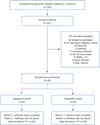
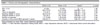
This is an ancillary study of a double-blinded, sham-controlled randomized clinical trial that investigated the effects of tDCS vs. sham-tDCS in patients with PSD.15 The study was unicentric, being conducted at the Hospital Universitário, Universidade de São Paulo, São Paulo, Brazil. We included 48 patients (Figure 1, Table 1), 24 in the tDCS and 24 in the sham-tDCS group. The primary outcome was score change in the 17-item Hamilton Depression Rating Scale at 6 weeks. Here, we investigated the effects of tDCS on cognitive performance trajectories among PSD patients as a secondary outcome of our trial.

| tDCS n=24 | Sham n=24 | t-test/?2 | p-value | |
|---|---|---|---|---|
| Sex, n (%) | 12 (50) | 12 (50) | ?2(1) = 0 | 1.00 |
| Age | 62.2 (12.3) | 61.3 (10.8) | t(44.68) = -0.27 | 0.79 |
| Years of education | 7.75 (5.7) | 7.57 (4.43) | t(43.18) = -0.12 | 0.90 |
| Baseline HDRS-17 score | 22.8 (3.14) | 21.2 (3.57) | t(45.25) = -1.63 | 0.11 |
| Endpoint HDRS-17 score | 13.2 (6.7) | 16.7 (4.44) | t(39.92) = 2.13 | 0.04 |
| Number of episodes | 1.17 (0.48) | 1.29 (0.62) | t(43.219) = 0.78 | 0.44 |
| Duration of the current MDD episode in weeks | 8.83 (8.11) | 19.6 (18.2) | t(28.73) = 2.54 | 0.02 |
Data presented as mean (SD), unless otherwise specified.
HDRS = 17-item Hamilton Depression Rating Scale; MDD = major depressive disorder; tDCS = transcranial direct current stimulation.
Participants
The patients were recruited through referrals from neurological and rehabilitation centers and from a naturalistic stroke surveillance cohort study. The inclusion criteria for the study were age 30 to 90 years, a 17-item Hamilton Depression Rating Scale score = 17, which indicated moderate to severe depression. Only individuals experiencing their first depressive episode were eligible, which had to begin 1 to 12 months after the stroke. The participants' first stroke had to have occurred in the past 5 years. Low suicide risk was required, which was determined by a score = 2 on the suicide item (the third item) of the 17-item Hamilton Depression Rating Scale. Finally, participants were required to have a DSM-IV diagnosis of "mood disorder due to a general medical condition (stroke) with a major depressive-like episode." The exclusion criteria were any other current Axis I disorder except anxiety disorders. Participants with specific contraindications for tDCS, such as metallic plates in the head, were also excluded. Other disqualifying conditions included neurological disorders (e.g., dementia and epilepsy) and life-threatening clinical conditions. Individuals taking benzodiazepines in doses exceeding 10 mg/day (or the equivalent) were excluded, as were those using any antidepressants, antipsychotics, sedatives, or hypnotics, although anticonvulsants were permitted if prescribed for previous stroke-related seizures (for details please refer to Valiengo et al.15). Stroke diagnosis was confirmed by a physician through history-taking, physical examination, and neuroimaging. Depression was diagnosed by a trained psychiatrist using the Mini-International Neuropsychiatric Interview for DSM-IV psychiatric disorders.
Interventions
Patients were randomly allocated to receive either active or sham-tDCS according to a randomized, sham-controlled, double-blind trial design with a 1:1 permuted block randomization. Raters, operators, and patients were all blinded to the treatment assignments. To further ensure blinding, contact between participants was minimized. Stimulation was administered using a standard tDCS device (DC-Stimulator, Neuroconn, Ilmenau, Germany) with a bifrontal electrode montage. The anode and cathode were placed over the left and right dorsolateral prefrontal cortex, respectively, at the F3 and F4 sites according to the international 10-20 electroencephalogram system. Rubber electrodes were placed in 25 cm2 saline-soaked sponges and were fastened with a headband. The current intensity was set at 2 mA. Participants remained seated at rest in a quiet, isolated room without external stimuli, and no specific tasks were assigned during the sessions.15 Twelve stimulation sessions (anodal left/cathodal right dorsolateral prefrontal tDCS, 30 min each, once daily on weekdays for 2 weeks, then one session every other week) were administered over 6 weeks. The endpoint was based on our previous study.20 To mimic the common skin sensations experienced immediately after stimulation, the sham-tDCS consisted of only 60 seconds of stimulation, followed by no stimulation for the remainder of the session. Randomization was performed with an automated device that assigned either sham or active stimulation based on a code. The codes were randomized by a research assistant who had no involvement in other aspects of the trial. These codes were then entered by the study nurse, who was blinded to the participants' group assignments.
Neuropsychological assessment
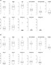
The neuropsychological battery was administered twice: on day 1, before the first stimulation session, and during week 6 (Figure 2 and Supplementary Material S1). The battery included: 1) the Montreal Cognitive Assessment; 2) the Mini-Mental State Examination (MMSE), both of which are used for cognitive screening21,22; 3) the Frontal Assessment Battery (FAB), which is designed to assess the cognitive and behavioral domains of executive function23; 4) the Trail Making Test, which evaluates processing speed, visual scanning, psychomotor activity, and overall executive function - scores are measured in seconds, with higher values indicating worse performance (the test is divided into two parts, the first involves connecting numbers in ascending order with a line, and the second requires alternating between numbers [in ascending order] and letters [in alphabetical order] with a line)24; 5) the Digit Span subtest of the Wechsler Adult Intelligence Scale, which assesses verbal short-term memory and working memory, cognitive control, and executive function - the subtest is divided into two parts: the first involves verbal repetition in forward order, and the second part involves verbal repetition in backward order)25; 6) the Wechsler Adult Intelligence Scale Digit Symbol-Coding task, which requires the reproduction of abstract symbols corresponding to associated numbers, with higher execution times indicating poorer performance26; 7) the Stroop Color and Word Test, which assesses the ability to suppress automatic responses. The test consists of three parts, with longer execution times indicating poorer performance. Time and errors are recorded.27

Cognitive domains
The neuropsychological tests were grouped into cognitive domains. A parallel analysis of standardized tests determined that the optimal number of factors was three. Exploratory factor analysis with oblique rotation was then conducted using combined baseline and endpoint data. Confirmatory factor analysis was performed using the lavaan package (Supplementary Figures S1, S2, and S3, and Supplementary Table S1) with the following domains: 1) "Stroop effect," which included cognitive control, attention, and inhibition of cognitive interference (Stroop color-naming, word-reading, and color-word tasks); 2) "processing speed" (Trail Making-A, Trail Making-B, and Digit Symbol-Coding); and 3) "executive function" (Digit Forward, Digit Backward, FAB). Raw scores were standardized (z scores) to create composite cognitive domain measures. Considering the importance of language and memory, we grouped the respective subtests of the Montreal Cognitive Assessment and MMSE. However, due to a high number of missing values and low variability of the subtests, the models did not converge and were not included.
Statistical analysis
The analysis was conducted in R 4.3.1. Results with p < 0.05 were considered significant. The number of comparisons was reduced by grouping cognitive domains. Extreme values (outside the 5th to 95th percentiles) in raw test scores were Winsorized. Trail Making-A and Trail Making-B results were multiplied by (-1). For the Stroop Test, rates (R) were calculated for the color-naming, word-reading, and color-word results, considering correct responses (C) and time (T) in seconds (RCN = CCN/TCN, RWR = CWR/TWR, RCW = CCW/TCW), based on Scarpina & Tagini.27 The rates were then multiplied by (-1).
The change in each cognitive test and domain was modeled using linear mixed models. Time, group, and their interaction were defined as fixed factors, while measurements were nested within the patients. A random effect for the intercept was included at the patient level. We refrained from imputing missing values, as our analysis assumed their randomness, and linear mixed models can accommodate this variability. The significance of the model factors was computed using Satterthwaite's approximation for degrees of freedom. Effect sizes were reported as Cohen's d.28,29
As in Moreno et al.,30 we included three different models for each cognitive test and domain, considering that age, sex, and education are known confounding factors that can impact depression and cognition: 1) unadjusted for covariates; 2) controlled for age, sex, and education; and 3) adjusted for age, sex, education, and change in depression (Hamilton Depression Rating Scale score post- vs pre-treatment).
We performed two general linear models to examine the relationships between baseline cognitive function and changes in depressive symptoms, as well as between baseline depression severity and changes in cognitive function, while considering the potential impact of the treatment group. Specifically, the first general linear model determined whether the initial cognitive state is associated with subsequent changes in depressive symptoms, considering treatment group and interaction. Conversely, the second general linear model determined whether the initial depressive state is associated with subsequent changes in cognitive function, also considering treatment group and interaction.
Ethics statement
Written informed consent was obtained from all patients prior to enrollment. The study was approved by the Hospitals' ethics committees (decisions 0062.0.198.000-12 and 76259023.3.0000.0068) and was registered with ClinicalTrials.gov NCT01525524.
RESULTS
Overview
Of almost 200 volunteers, we included 48 patients who met inclusion criteria (Figure 1 15 and Table 1). There were no significant differences in clinical or demographic characteristics between the groups, except for the duration of the current episode, which was longer in the sham-tDCS group.
Results of individual cognitive tests
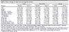
Significant time × group interactions were found for the FAB (model 2 F1,39.46 = 5.238, p = 0.027, and model 3 F1,39.46 = 5.19, p = 0.028, both d = -0.69, 95%CI -1.28 to -0.10) and MMSE (model 2 F1,39.98 = 4.121, p = 0.049, d = 0.69, 95%CI 0.09 to 1.27 and model 3 F1,39.91 = 4.147, p = 0.048, d = 0.69, 95%CI 0.09 to 1.28). Digit Span forward showed trend results (Supplementary Table S2). The group mean differences suggested that the sham-tDCS group performed better in the FAB and in the active group performed better in the MMSE. Both groups showed improvement in the Digit Span forward task, although the difference was only significant for the sham-tDCS group (Table 2). Significant within-group differences were also found for the sham-tDCS group in FAB, Digit Span forward, and Trail Making-A, and significant within-group differences were found in the tDCS group for the Montreal Cognitive Assessment, Stroop word-reading task, Stroop color-word task, and Stroop effect z score (Table 3).
| Measure | Model | Sham-tDCS vs. tDCS | ||
|---|---|---|---|---|
| Difference | p-value | Effect size | ||
| Stroop effect | 1 | 0.16 (0.15) | 0.305 | 0.38 (-0.31 to 1.08) |
| 2 | 0.11 (0.16) | 0.485 | 0.28 (-0.42 to 0.97) | |
| 3 | 0.11 (0.16) | 0.515 | 0.26 (-0.44 to 0.95) | |
| Processing speed | 1 | -0.22 (0.12) | 0.100 | -0.51 (-1.42 to 0.42) |
| 2 | -0.24 (0.14) | 0.111 | -0.54 (-1.45 to 0.39) | |
| 3 | -0.24 (0.14) | 0.109 | -0.54 (-1.45 to 0.38) | |
| Executive function | 1 | -0.18 (0.11) | 0.135 | -0.45 (-1.05 to 0.15) |
| 2 | -0.24 (0.11) | 0.035 | -0.62 (-1.23 to -0.01) | |
| 3 | -0.24 (0.11) | 0.035 | -0.62 (-1.23 to -0.01) | |
Bold type denotes statistical significance.
tDCS = transcranial direct current stimulation.
Model 1 was an unadjusted linear mixed model with time, group, and their interaction as fixed factors; Model 2 additionally included age, sex, and education; Model 3 included time and 17-item Hamilton Depression Rating Scale depression scores as a covariate; Cohen's d; effect sizes are shown with 95%CI in parenthesis; the numbers are rounded to two decimal points.
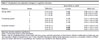
| Measure | tDCS | Sham-tDCS | Change | |||
|---|---|---|---|---|---|---|
| Baseline | Week 6 | Baseline | Week 6 | tDCS | Sham | |
| FAB | 11.60 (2.90) | 11.40 (3.37) | 9.33 (3.02) | 11 (2.96) | -0.25 (2.73) | 1.19 (3.12)* |
| MOCA | 18.10 (3.27) | 19.50 (3.71) | 17.20 (3.61) | 18.40 (2.44) | 1.30 (2.52)* | 0.75 (2.62) |
| MMSE | 23.30 (3.24) | 24.40 (3.79) | 23.60 (3.02) | 23 (3.38) | 1.05 (3.23) | -1.10 (3.49) |
| Digit Span - forward | 6.32 (1.85) | 6.48 (1.82) | 5.25 (1.77) | 6.19 (1.28) | 0.13 (1.66) | 1.02 (1.69)* |
| Digit Span - backward | 3.52 (2.06) | 3.19 (1.47) | 3.55 (2.36) | 3.04 (1.49) | -0.43 (1.77) | -0.80 (2.56) |
| Digit Symbol-Coding | 2.45 (4.53) | 0.92 (1.81) | 0.94 (1.95) | 1.24 (1.98) | -1.76 (3.89) | 0.24 (3.12) |
| Trail Making-A | -123 (84.2) | -103 (50.5) | -141 (83.7) | -111 (56.4) | 2.29 (39.3) | 23.80 (59.2)* |
| Trail Making-B | -312 (239) | -213 (150) | -313 (169) | -324 (160) | 26.90 (69.9) | 13.00 (173) |
| Stroop CN | -8.48 (18.2) | -1.19 (0.43) | -7.66 (18) | -1.08 (0.32) | 8.67 (20) | 6.90 (18.6) |
| Stroop WR | -2.68 (2.05) | -1.62 (0.78) | -2.41 (1.53) | -1.79 (0.46) | 0.84 (1.69)┼ | 0.22 (1.18) |
| Stroop CW | -4.25 (3.23) | -2.85 (1.58) | -3.13 (2.17) | -2.74 (1.34) | 1.20 (2.53)┼ | -0.09 (1.11) |
| Stroop effect z score | 0.01 (2.63) | 1.32 (0.88) | 0.63 (1.53) | 1.18 (0.68) | 1.21 (2.50)┼ | 0.17 (0.99) |
| Processing speed z score | 0.51 (2.72) | 0.71 (1.66) | -0.23 (1.55) | 0.40 (1.45) | -0.47 (1.08) | 0.87 (1.19) |
| Executive function z score | 0.64 (2.24) | 0.52 (2.09) | -0.60 (1.60) | 0.17 (1.81) | -0.31 (1.93) | 0.55 (1.77) |
CN = color-naming; CW = color-word; FAB = Frontal Assessment Battery; MMSE = Mini-Mental State Examination; MOCA = Montreal Cognitive Assessment; tDCS = transcranial direct current stimulation; WR = word-reading.
The mean and SD are shown for each group at baseline and week 6. Change in scores was calculated as the score at baseline minus the score at 6 weeks, with greater increases indicating better cognitive functioning. Trail Making-A, Trail Making-B, and Stroop color-naming, Stroop word-reading, and Stroop color-word results were multiplied by (-1), i.e., higher values indicate better performance.
* Significant within-group change.
Cognitive domain results
Significant time × group interactions were found for executive function in adjusted models 2 and 3 (F1, 38.36 = 4.76, p = 0.035, d = -0.62, 95%CI -1.23 to -0.01). The group mean differences indicated slight worsening in the active-tDCS group and slight improvement in the sham-tDCS group. The groups did not differ significantly at the endpoint. No significant effects were found for Stroop effect and processing speed (Table 2).
Associations with improved cognitive function
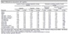
A significant interaction effect was found between group and depressive symptoms for the Stroop color-naming task (ß = -5.657, standard error = 1.745, t(30) = -3.241, p = 0.003, positive false discovery rate = 0.006) and the Stroop color-word task (ß = -0.448, standard error = 0.180, t(26) = -2.490, p = 0.020, positive false discovery rate = 0.039) (Table 4 and Supplementary Figure S4). Improvement in the Stroop color-naming task was associated with worse depressive scores at baseline for the sham-tDCS group, whereas the opposite pattern occurred in the tDCS group, with improvement linked to lower Hamilton Depression Rating Scale scores. Stroop color-word scores in the sham-tDCS group did not change in relation to baseline Hamilton Depression Rating Scale scores, while lower Hamilton Depression Rating Scale scores in the tDCS group were associated with better performance. A trend towards a significant interaction effect was observed between group and depressive symptoms for Trail Making-B (ß = -37.233, standard error = 17.332, t(16) = -2.148, p = 0.047, positive false discovery rate = 0.054). This interaction suggested that improvement in Trail Making-B was more affected by baseline depression scores in the sham-tDCS group.
| Cognitive function predicting depression change (HDRS) | Depression predicting cognitive function change | |||||||
|---|---|---|---|---|---|---|---|---|
| Cognition | Cognition × Group | Depression symptom | Depression symptom × Group | |||||
| Measures | F-value | p-value | F-value | p-value | F-value | p-value | F-value | p-value |
| MOCA | 0.57 | 0.45 | < 0.01 | 0.99 | 0.09 | 0.76 | 0.11 | 0.74 |
| MMSE | 0.11 | 0.74 | 0.81 | 0.37 | 2.09 | 0.16 | 0.23 | 0.59 |
| FAB | 0.69 | 0.41 | 0.20 | 0.66 | 0.53 | 0.47 | 0.05 | 0.83 |
| Digit Span - forward | 2.00 | 0.16 | 0.47 | 0.50 | 0.45 | 0.50 | 0.10 | 0.76 |
| Digit Span - backward | 0.28 | 0.60 | 0.013 | 0.91 | 0.23 | 0.63 | 1.49 | 0.23 |
| Digit Symbol-Coding | 1.70 | 0.20 | 0.03 | 0.86 | 0.38 | 0.54 | 0.81 | 0.38 |
| Trail Making-A | 0.88 | 0.35 | 0.11 | 0.74 | 0.55 | 0.46 | 0.15 | 0.70 |
| Trail Making-B | <0.01 | 0.96 | 0.07 | 0.79 | 1.65 | 0.29 | 4.73 | 0.05 |
| Stroop CN | 0.85 | 0.36 | 0.27 | 0.60 | 1.10 | 0.30 | 10.53 | < 0.01 |
| Stroop WR | 0.55 | 0.46 | 0.14 | 0.71 | 0.87 | 0.36 | 3.07 | 0.09 |
| Stroop CW | 2.14 | 0.15 | 0.05 | 0.82 | 4.42 | 0.04 | 6.21 | 0.02 |
| Stroop effect z score | 1.49 | 0.23 | 0.31 | 0.58 | 3.47 | 0.07 | 4.07 | 0.05 |
| Processing speed z score | 0.93 | 0.35 | 0.24 | 0.63 | 0.07 | 0.80 | 3.13 | 0.11 |
| Executive function z score | 1.30 | 0.26 | 0.11 | 0.74 | 0.01 | 0.91 | 0.03 | 0.86 |
CN = color-naming; CW = color-word; FAB = Frontal Assessment Battery; HDRS = Hamilton Depression Rating Scale; MMSE = Mini-Mental State Examination; MOCA = Montreal Cognitive Assessment; WR = word-reading.
Numbers rounded to two decimals; significance of model factors computed using analysis of variance.
Bold type indicates significant difference.
DISCUSSION
In this ancillary study of Valiengo et al.15 on the efficacy of tDCS for PSD, we explored the effects of tDCS in the cognitive domains of Stroop effect, processing speed, and executive function, analyzing several individual tasks. The study's strengths included its effective blinding and antidepressant-free sample, since antidepressant drugs can interfere with cognition. Our findings revealed no consistent evidence that tDCS significantly improved cognitive domain performance, although in individual tasks active tDCS led to Mini-Mental State Examination improvement compared to sham-tDCS, while sham-tDCS treatment led to FAB improvement.
Specifically, in the domain of executive function, we observed slight worsening in the active-tDCS group and slight improvement in the sham-tDCS group. The association between PSD and cognitive impairment has been well-established for executive dysfunction.31 Corroborating this finding, a meta-analysis of 27 studies targeting the dorsolateral prefrontal cortex revealed that tDCS did not significantly improve general executive function.18 Interestingly, active tDCS led to greater improvement in Mini-Mental State Examination individual task scores than sham-tDCS. The Mini-Mental State Examination is a widely used screening test for cognitive impairment and dementia in both clinical and research settings. The test consists of a short battery that includes subitems evaluating orientation, short memory, attention, and language. Although this instrument is generally useful for investigating dementia and mild cognitive impairment, previous studies have shown that post-stroke patients with PSD had significantly lower Mini-Mental State Examination scores than non-depressed post-stroke patients with comparable lesion locations and volume.32 This suggests tDCS may assist in general cognitive function. On the other hand, the sham-tDCS group's improvement in FAB scores may be due to practical effects, i.e., since it is a brief cognitive test, the participants could become familiarized with the tasks through repeated exposure, leading to better performance despite no actual treatment effect.
A bidirectional association with cognition that investigated 1) whether cognitive function predicted change in depression and 2) whether depression predicted change in cognitive function showed significant results for the latter regarding the Stroop color-naming task and the Stroop color-word task, as well as a trend toward significance in Trail Making-B results. For the Stroop color-naming task, a congruent subtest in which participants name the color of inked squares, the active-tDCS group with less severe depression (according to Hamilton Depression Rating Scale scores) performed better, while improvement in the sham-tDCS group was associated with more severe depression. However, for the more complex Stroop color-word subitem, which requires patients to name the color in which a word is printed, rather than the word itself (thus inhibiting an automatic response), improvement following tDCS was associated with higher Hamilton Depression Rating Scale scores. This suggests a distinct pattern of interaction between the dorsolateral prefrontal cortex and tDCS, with depression severity influencing cognitive processing differently. It may indicate that varying cognitive mechanisms or neural pathways are influenced by task complexity and depression level, resulting in different effects from tDCS on performance.
Cognitive improvement after tDCS involves significant intra- and inter-subject variability when used for stroke treatment, likely due to the specific brain regions affected by the stroke and the extent of impairment.33,34 Another crucial factor is the timing of tDCS therapy. The acute and subacute stages after stroke are ideal for stimulation, with the greatest improvement in cognition typically occurring within the first 6 months.35 However, depression can occur up to 12 months after a stroke, thus missing the optimal treatment window. Additionally, the duration of depressive episode in our sample was relatively long, and only 16.6% of our sample was treating depression with antidepressants prior to the drug washout. The duration, severity, and untreated state of depression can significantly exacerbate cognitive dysfunction.36
This study has certain limitations that should be noted. Because cognition was an ancillary outcome of the main trial, the tDCS treatment parameters and sample size might not have been adequate to affect cognition. Moreover, the multiple analysis of cognitive outcomes could have led to false positive results, so the data must be interpreted accordingly. A further limitation is that we did not control the patients' cognitive profile according to stroke location. Different topographical lesions can lead to different deficits in cognitive functioning. Another possible limitation is that we did not consider the effects of circadian rhythm or sleep pressure. Both could affect cognition and tDCS-induced neuroplasticity.37-39 Although all cognition measurements were collected at the same time of the day, we did not control for sleep pressure.
In conclusion, this ancillary study found no consistent evidence that tDCS significantly improved cognitive performance in broad domains like the Stroop effect, processing speed, or executive function in patients with PSD. While the active-tDCS group improved in MMSE results, the sham-tDCS group improved in FAB results, likely due to practice effects. A key finding was slight worsening in executive function in the active-tDCS group compared to slight improvement in the sham group, which aligns with previous findings that tDCS does not significantly affect general executive function. According to the bidirectional analysis of depression and cognition, depression severity affected cognitive task performance differently in the active-tDCS and sham groups. In the Stroop color-naming task, patients with less severe depression showed greater improvement in the active-tDCS group, while in the more complex Stroop color-word task, higher depression severity was linked to improvement after active-tDCS. These findings highlight the complexity and variability of cognitive improvement after tDCS, which may be influenced by depression severity and task complexity. Additionally, the timing of tDCS in relation to stroke recovery is critical, since treatment during the acute and subacute phases may yield better cognitive outcomes.
SUPPLEMENTARY MATERIALS
ACKNOWLEDGMENTS
This study was supported by a research grant to AG from Fundação de Amparo à Pesquisa do Estado de São Paulo (FAPESP; grant 2011/22872-4; 2012/20911-5), which is an independent public foundation and had no role in any aspect of the study, including design and conduct of the study, the collection, management, analysis and interpretation of the data, or the preparation, review, or approval of the manuscript. TZ was supported by FAPESP (grant 2020/03235-2), BSP was supported by FAPESP (grant FAPESP 2022/15368-2), RRV was supported by Coordenação de Aperfeiçoamento de Pessoal de Nível Superior (Social demand).
 PDF
 Print
 Send this article by email
 How to cite this article
 Submit a comment
 Mendeley
 Pocket
 Share on Facebook
 Share on Twitter